Thalassemias are a group of blood disorders that affect the way
the body makes hemoglobin, a protein found in red blood cells that is
responsible for carrying oxygen throughout the body.
The body
contains more red blood cells than any other type of cell, and each has a
life span of about 4 months. Each day, the body produces new red blood
cells to replace those that die or are lost from the body.
With a
thalassemia, the red blood cells are destroyed at a faster rate,
leading to anemia, a condition that can cause fatigue and other
complications.
Thalassemias are inherited conditions — they're
carried in the genes and passed on from parents to children. People who
are carriers of a thalassemia gene show no thalassemia symptoms and
might not know they're carriers. If both parents are carriers, they can
pass the disease to their kids. Thalassemias are not contagious.
While there are many different types of thalassemias, the main two are: Alpha thalassemia: when the body has a problem producing alpha globin Beta thalassemia: when the body has a problem producing beta globin
When
the gene that controls the production of either of these proteins is
missing or mutated, it results in that type of thalassemia. About Alpha Thalassemia
Alpha
thalassemia occurs when the gene that controls the making of alpha
globins is absent or defective. It can be mild to severe and is most
commonly found in people of African, Middle Eastern, Chinese, Southeast
Asian, and, occasionally, Mediterranean descent.
Some children
with alpha thalassemia have no symptoms and may require no treatment.
Others with more severe cases need regular blood transfusions to treat
anemia and other symptoms.
A child can only get alpha thalassemia
by inheriting it from his or her parents. Genes are "building blocks"
that play an important role in determining physical traits and many
other things about us.
Humans are made up of trillions of cells
that form the structure of our bodies and carry out specialized jobs
like taking nutrients from food and turning them into energy. Red blood
cells, which contain hemoglobin, deliver oxygen to all parts of the
body.
All cells have a nucleus at their center, which is kind of
like the brain or "command post" of the cell. The nucleus directs the
cell, telling it to grow, mature, divide, or die. The nucleus contains
DNA (deoxyribonucleic acid), a long, spiral-shaped molecule that stores
the genes that determine hair color, eye color, whether or not a person
is right- or left-handed, and many more traits. DNA, along with genes
and the information they contain, is passed down from parents to their
children during reproduction.
Each cell has many DNA molecules,
but because cells are very small and DNA molecules are long, the DNA is
packaged very tightly in each cell. These packages of DNA are called
chromosomes, and each cell has 46 of them. Each package is arranged into
23 pairs — with one of each pair coming from the mother and one from
the father. When a child has alpha thalassemia, there is a mutation in
chromosome 16.
Alpha globin is made on chromosome 16. So, if any
gene that tells chromosome 16 to produce alpha globin is missing or
mutated, less alpha globin is made. This affects hemoglobin and
decreases the ability of red blood cells to transport oxygen around the
body.
Alpha thalassemia refers to a group of disorders characterized by
inactivation of alpha globin genes. This results in a relative increase
in nonfunctional beta globin or gamma globin tetramers and subsequent
cell damage. Normally, there are four alpha genes. Absence or
non-function of three alpha genes results in hemoglobin H disease, and
the loss of all four alpha genes usually results in intrauterine death.
The alpha thalassemias are caused by a decrease in production of alpha
globin chains due to a deletion or mutation of one or more of the four
alpha globin genes located on chromosome 16. Alpha gene mapping can be
obtained to determine the specific mutation. The alpha thalassemias can
be generally categorized as: Silent Carrier, Alpha Thalassemia Trait,
Hemoglobin H disease, Hemoglobin H-Constant Spring, and Alpha
Thalassemia major. Frequently, the diagnosis of alpha thalassemia trait
in a parent is discovered after the birth of an affected child.
The Silent Carrier status is characterized by three functional genes
that code for the production of alpha globins (-a/aa). Outside the
newborn period, it is not possible to make this diagnosis by
conventional methods. There is overlap between the red blood cell
indices of these individuals and normals, although the MCV may be
slightly lower. The silent carrier will experience no health problems in
his/her lifetime. This carrier state is diagnosed by deduction when a
'normal' individual has a child with Hgb H disease or with microcytic
anemia consistent with alpha thalassemia trait. An unusual case of the
silent carrier state is the individual who carries the Hemoglobin
Constant Spring mutation [(acsa/aa) or (aacs/aa)]. This is an elongated
a-globin due to a termination codon mutation. Individuals who have this
mutation have normal red blood cell indices, but can have children who
have Hgb H-Constant Spring disease if the other parent has alpha
thalassemia trait (--/aa). Generally, children with Hgb H-Constant
Spring are more affected clinically than children who have classic Hgb H
disease. Two Constant Spring carriers can also pass on their genes to
have a child with Homozygous Constant Spring, a condition that has
similar clinical implications as Hemoglobin H disease.
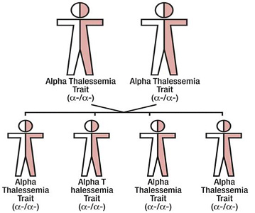
Alpha thalassemia trait is characterized by two functional genes that
code for the production of alpha globins [(-a/-a) or (--/aa)]. The two
genes can either occur on the same chromosome (cis-type) or on each of
the pair (trans-type). Cis-type a-thalassemia trait tends to be found in
individuals of Asian descent, while trans-type tends to run in
individuals of African descent. Cis-type can be co-inherited with
another cis-type or hemoglobin H disease to result in alpha thalassemia
major, or hydrops fetalis. Individuals who have alpha thalassemia trait
are identified by microcytosis, erythrocytosis, hypochromia, and mild
anemia. The diagnosis is made by a combination of family studies and the
ruling out of both iron deficiency anemia and beta thalassemia trait.
In the neonatal period, when hemoglobin Bart's (g4) is present, the
diagnosis can be strongly suspected. In children, there are no markers
such as Hgb A2 and Hgb F to make the diagnosis. (One exception is the
case where both of the deletions occur on the same chromosome and zeta
[z] globin is expressed in carriers. This is most common in Southeast
Asians.) The diagnosis is one of exclusion. The clinician should be
satisfied with the presumed diagnosis if the above criteria are met.
During pregnancy, the microcytic anemia can be mistaken for anemia of
pregnancy.
The individual with a thalassemia trait will experience no significant
health problems except a possible slight anemia which cannot be treated
with iron.
Hemoglobin H disease is characterized by one functional gene that codes
for the production of alpha globins (--/-a). Hgb H disease should be
considered in the case of a neonate in whom all of the red blood cells
are very hypochromic. These neonates have a high percentage of
hemoglobin Bart's on the their newborn screening results. In older
children, this hemoglobinopathy is characterized by moderate anemia with
a hemoglobin in the 8 to 10 gm/dL range, hypochromia, microcytosis, red
cell fragmentation, and a fast migrating hemoglobin (Hgb H) on
electrophoresis.
Hemoglobin H does not function as a normal hemoglobin and has a high
oxygen affinity (holds onto oxygen longer making it unavaible for use by
the body), so the measured hemoglobin in these children is misleading.
Individuals who have Hgb H generally have a persistent stable state of
anemia, which may be accentuated by increased hemolysis during viral
infections and by exposure to oxidant medications, chemicals and foods
such as sulfa drugs, benzene, and fava beans (similar to individuals who
have G6PD deficiency). As the red cells mature they loose their ability
to withstand oxidant stress and Hgb H precipitates, leading to
hemolysis. Therapy for individuals who have Hgb H disease includes
folate, avoidance of oxidant drugs and foods, genetic counseling
education and frequent medical care. Uncommon occurrences in a child
with Hgb H would be severe anemia, skin ulceration, and splenomegaly
requiring splenectomy. Unlike individuals who have beta thalassemia,
hemosiderosis is rare in Hgb H disease.
Children with Hemoglobin H-Constant Spring have a more severe course
than children who have Hgb H. They have a more severe anemia, with a
steady state hemoglobin ranging between 7 and 8 gm/dl. They more
frequently have splenomegaly and severe anemia with febrile illnesses
and viral infections, often requiring transfusion. If anemia is
chronically severe and the child has splenomegaly, a splenectomy may be
performed. If splenectomy is anticipated, the complication of severe
post-splenectomy thrombocytosis with hypercoagulability can occur,
leading to thrombosis of the splenic vein or hepatic veins. This
complication has also been reported as recurrent pulmonary emboli and
clotting diathesis. At Children's Hospital Oakland, children who are
scheduled to have surgery are treated pre-surgically with low molecular
weight heparin, followed by low dose aspirin, continued indefinitely.
The most severe form of alpha thalassemia is Alpha Thalassemia Major or
hydrops fetalis, characterized by a deletion of all four genes that
code for alpha globins . This diagnosis is frequently made in the last
months of pregnancy when fetal ultrasound indicates a hydropic fetus.
The mother frequently exhibits toxemia and can develop severe postpartum
hemorrhage. These infants are usually stillborn. There can be other
congenital anomalies, though none are pathognomonic for alpha
thalassemia major. Since alpha globins are required for production of
fetal and adult hemoglobin, the fetus suffers from signifiant in utero
hypoxia. The only hemoglobins found in these infants are: Hgb Portland
(d2g 2), Hgb H (b), and Hgb Bart's (g4), and no Hgb A or A2. These
babies can have other complications associated with hydrops, such as
heart failure and pulmonary edema.
If the diagnosis is made early, intrauterine transfusions can be
performed. There are reports of survival and chronic transfusion in
these infants; CHO cares for one alpha thalassemia major baby with
chronic transfusion therapy. Undoubtedly, more of these infants could be
saved if the diagnosis is anticipated by prenatal diagnosis and
treatment provided.